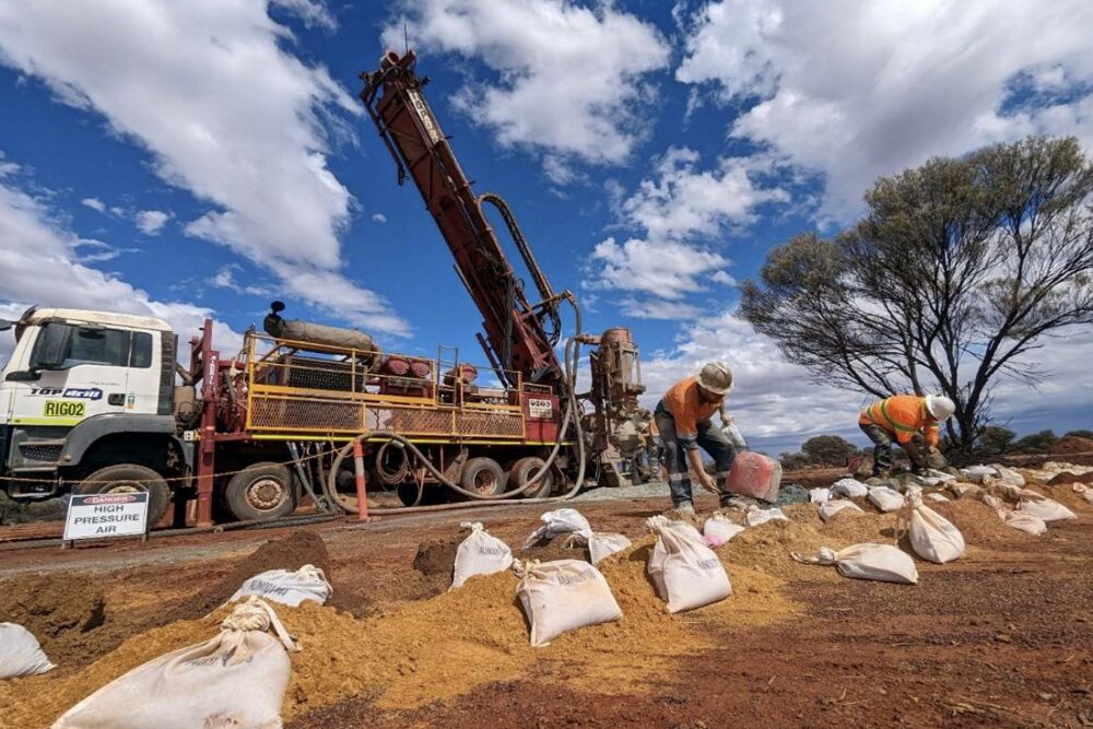


The mining industry is set to benefit from a new Australian capability that uses a nuclear scanning technique to detect the presence of precious metals and strategic minerals in a core sample.
The approach, which involved enhancements to the neutron tomography instrument Dingo at the Australian Nuclear Science and Technology Organisation’s (ANSTO) Australian Centre for Neutron Scattering, is expected to be available to industry later in the year.
A neutron tomography scan is similar to an X-ray CT scan but uses neutrons, neutral subatomic particles, produced by the OPAL multipurpose reactor.
Neutrons provide an alternative and complementary contrast to X-rays, in essence, a ‘fresh set of eyes’ on the same material.
“Traditionally, neutron CT scans took much longer than X-ray CT scans. With this new development, it has become a fast, cost-effective and non-destructive way to map the concentration and distribution of minerals in a rock core,” shared instrument scientist Dr Joseph Bevitt, who worked with a team at ANSTO and Macquarie University to add the capability to the instrument.
The concept was tested successfully with the support of Aurelia Metals Ltd. who provided cores for testing from the Hera Mine, a gold-lead-zinc-silver deposit located 100 kilometres south-east of Cobar in NSW, and in collaboration with Dr Timothy Murphy from Macquarie University’s Geoanalytical laboratory.
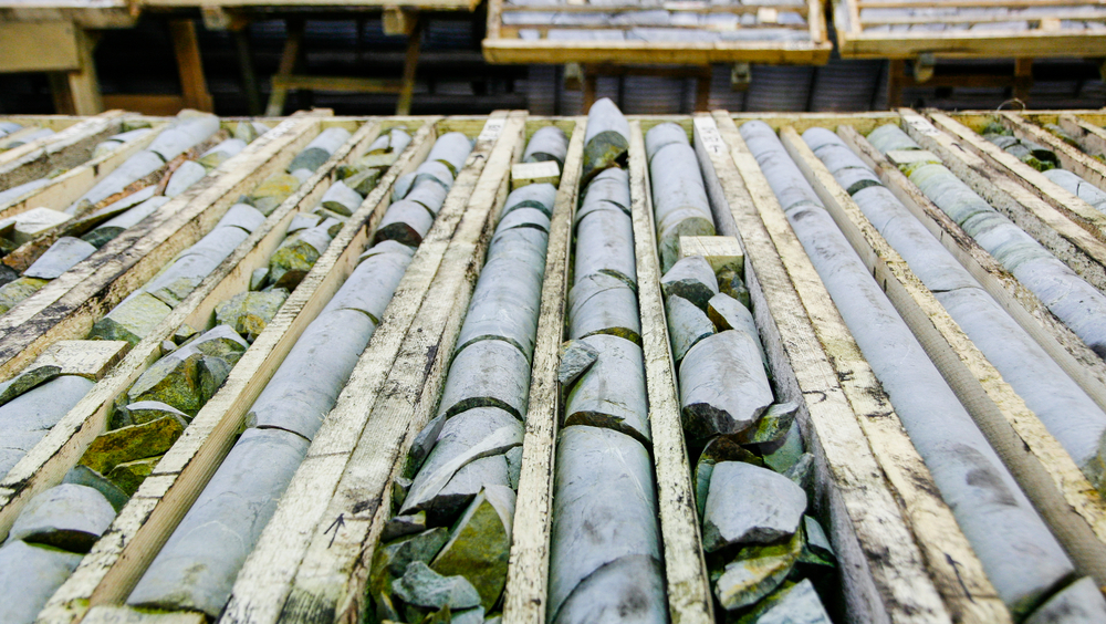
The ANSTO team comprising engineering and technical staff, developed a four-row rig, to hold the cores for parallel scanning.
The instrument produces a three-dimensional image reconstruction of the drillcore, which is achieved by rotating the cores in the neutron beam while acquiring thousands of shadow radiographs.
Using high-powered computing facilities, these are converted into 3-D visualisations of the drillcores. This data can be used to extend 2-D surface mineral maps achieved using X-ray fluorescence to more accurately report mineral content within entire drillcores.
“Our collaborators at Macquarie University were satisfied that the images were viable for mineralogical assessments and are developing new methods to explore and integrate these datasets into geological and geochemical analyses,” said Dr Bevitt.
Depending on the desired scan resolution, non-destructive neutron CT scanning of one- metre drillcore lengths can be completed in an hour.
At present, industry and research institutions use X-ray techniques for high throughput drillcore inspection and analysis. However, X-rays cannot penetrate samples that contain abundant heavy metals, such as lead, without losing image contrast.
“Neutrons overcome this limitation as lead, and a number of other commonly occurring minerals that are problematic for X-rays are more transparent to neutrons,” said Dr Bevitt.
The cartridges in the scanning rig can hold four cores up to 1.5 metres in length, with a maximum diameter of 80 mm each.
The team intends to make further improvements upon the prototype apparatus, including increased parallelisation to increase output by 50 per cent and robotic changeover of sample cartridges for automated bulk scanning studies.
Most importantly, planning is underway for the installation of an X-ray source to enable bi-modal neutron and X-ray tomographic imaging of drillcores.
Dr Bevitt said bi-modal imaging opens up the opportunity for holistic 3D mineral mapping, based on the combination of independent X-ray and neutron contrasts.
“This is a new application, as our instrument is used primarily for the analysis of cultural heritage materials, palaeontological specimens, and engineering materials.”
“Given the importance of the mining industry in Australia, we think this new technique will enhance exploration activity, and better inform environment sustainability,” he added.